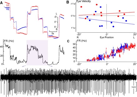
Fig. 1.

Conjugate left side horizontal eye position neural integrator (HPNI) neuron during spontaneous fixation behavior in darkness. A: FR (firing rate) in Hz correlated with left (LE, red) and right (RE, right) eye position following saccades of different amplitude. Inset: activity is expanded below as indicated by the arrows. B: P-V (eye position-velocity) plot constructed from only the fixations shown in A. Eye velocity (ordinate) was calculated from least squares regression of the eye position (filled part for each fixation). The time constant (τ) for LE was 159.7 s and RE 72.9 s. C: FR vs. eye position plot showing a correlation with both left and right eye position. Averaged LE and RE position and velocity coefficients were 1.59 (spike/s)/° and −0.29 (spike/s) per °/s, (r = 0.96) with + values leftward and – rightward.
