
Fig. 6.
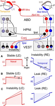
Schematic showing monocular fixation plasticity results and hypothesized neuronal connections. A–C: P (performance, dashed lines) and m (memory, solid lines) vignettes of monocular changes in time constant after 4 h visual training. Color-coding associates fixations in the LE and RE with the medial and lateral rectus eye muscles that can then be correlated with eye specific neurons in the vestibular nucleus (VEST), Area I (HPNI), and abducens (ABD) nuclei. A: RE instability and LE stability. B: RE leak and LE stability. C: RE leak and LE instability. In the diagram, monocular signaling is suggested for ipsilateral and conjugate eye movements with excitation (instability) originating from the left vestibular nucleus and inhibition (leak) from the right vestibular nucleus. Right inhibitory HPNI neurons (black) are shown to coordinate the proposed nasal and temporal eye position integrators. For simplification, connections are only shown to and from the right HPNI with direct vestibuloocular pathways to MR and Abd Mns omitted (see Supplemental Fig. S1A).