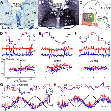
Fig. 7.

Integrator stability and time constant plasticity after acute midline lesion. A–C: anatomy of midline lesions between the bilateral HPNI nuclei. A: coronal photomicrograph showing an acute midline lesion encompassing ∼75% of the dorsal-ventral depth for behavior with P-V plots shown in D–H. B: 15 days post midline lesion photomicrograph showing extensive biocytin labeled axons in the medial longitudinal fasiculus (MLF) and reticular formation after spinal cord label. C: schematic illustrating 3 groups (a–c) of midline lesions (8 cases). D–F: P-V plots of eye position holding before (A) and 10 (B) and 30 min (C) after the midline lesion illustrated in A. Control LE τ was −127.8 s and RE −170.3 s in (A), −19.8 s and −16.6s in B, and 1,428.5 s and −384.3 s in C, respectively. G and H: vestibuloocular reflex (VOR) at 0.125 Hz and 15.7°/s before and 20 min after the lesion with least square regression fits of eye velocity. Head velocity shown in black. Gains changed minimally from (LE) 0.90 to 0.80 and (RE) 0.73 to 0.71 with negligible shifts in phase (LE) 3.2° to 2.3° and (RE) 0.2° to 3.0°. VNI, velocity neural integrator; FL, facial lobe; Vag, vagal lobe; VO, descending octaval nucleus; Xth N, vagus nerve; IO, inferior olive.
