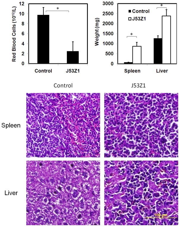
Figure 7. Development of erythroleukemia phenotypes in mice receiving implantation of J53Z1 cells.

Cultured J53Z1 cells (1×10⁶) were implanted into 12-week-old wild type C57Bl/6 mice through retro-orbital injections. Upper panel. Red blood cells, spleen, and liver were analyzed 2 to 4 weeks after implantation. Error bars denote standard deviation (n≥4), *P<0.001. Lower panel. Paraffin sections of spleen and liver from representative control and J53Z1-implanted mice were subjected to H&E staining. Note the loss of normal tissue architecture and infiltration of densely stained erythroleukemia cells in tissues from J53Z1-transplanted mice. Photos were taken with a 40x objective lens.
