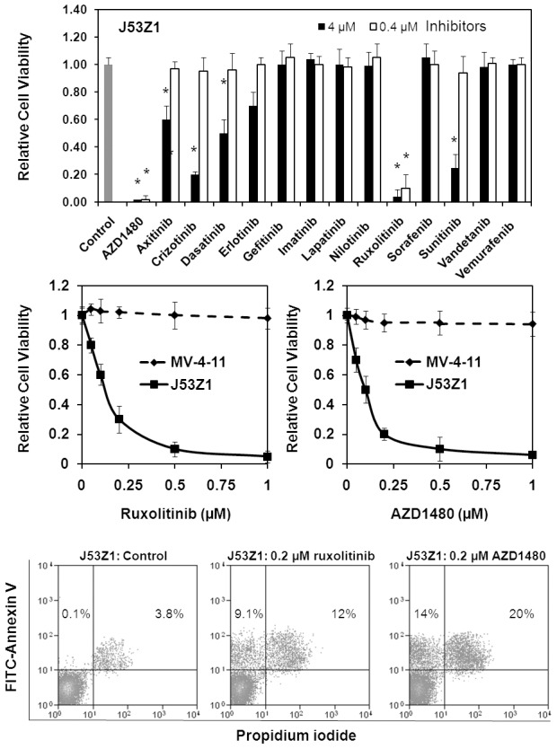
Figure 8. Inhibition of J53Z1 cells by selective protein kinase inhibitors.

J53Z1 cells were cultured in the presence of various concentrations of indicated protein kinase inhibitors. Top and middle panels. Cell viability was assessed by XTT assays after 72 hr of incubation. Control experiments were performed in the presence of 0.1% DMSO. Error bars denote standard deviation (n = 3). *P<0.001 in reference to control. Note that MV-4-11 cells were analyzed for comparison (middle panel). Bottom panel. Apoptosis assays were performed with J53Z1 cells after 24 hr of incubation with 0.2 µM of ruxolitinib or AZD1480. Cells were stained with FITC-annexin V and propidium iodide. Percentages of annexin V-positive cells are indicated.
