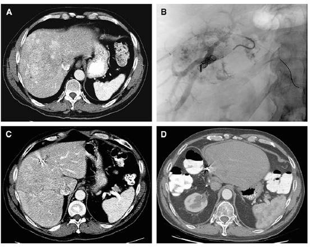
Figure 3.
A 59-year-old male patient had a 16-cm hepatocellular carcinoma in the right liver without evidence of extrahepatic disease. (A) Computed tomography revealed that the standardized future liver remnant (sFLR) volume was 12%.
(B) Right portal vein embolization (PVE) was performed. (C) Four weeks after PVE, the sFLR was 21%. (D) The patient had no evidence of disease 6 years post-resection. (Reprinted with permission from: Palavecino M, Chun YS, Madoff DC, et al. Surgery 2009;145:399–405)

