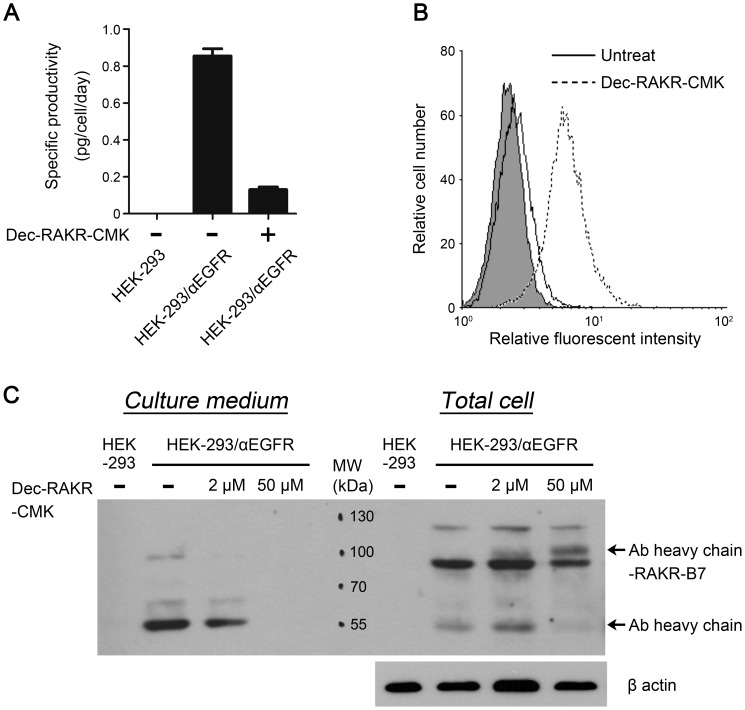
Figure 2. Furin inhibitor-mediated switch of secreted αEGFR Ab and anchored αEGFR Ab-RAKR-B7.
(A) HEK-293/αEGFR cells were treated with 20 µM furin inhibitor for 24 h, and HEK-293 cells were used as a negative control. Cultured medium was coated on 96-well plates and were detected by ELISA using HRP conjugated goat anti-human IgGFcγ antibody. Bars, SD. (B) HEK-293 cells (filled histogram); HEK-293/αEGFR cells (solid line) and HEK-293/αEGFR cells treated with furin inhibitor (20 µM) (dashed line) were analyzed by flow cytometry using a specific antibody to the human IgGFcγ to assess the expression of membrane-anchored αEGFR Ab-RAKR-B7. (C) The cultured medium and cell lysate harvested from HEK-293 cells, HEK-293/αEGFR cells, and HEK-293/αEGFR cells treated with furin inhibitor (2 or 50 µM) were analyzed by western blotting using HRP conjugated goat anti-human IgGFcγ antibody. The clear bands appearing at 55 KD in lanes 2 and 3 correspond to the secreted αEGFR Ab heavy chain, and the clear bands appearing at 95 KD in lanes 7 and 8 correspond to the membrane-anchored αEGFR Ab heavy chain-RAKR-B7. The bands appearing at 80 KD in lanes 7, 8, and 9 correspond to the αEGFR Ab light chain-2A-heavy chain, and the bands appearing at 120 KD in lanes 7, 8, and 9 correspond to the αEGFR Ab light chain-2A-heavy chain-RAKR-B7.

