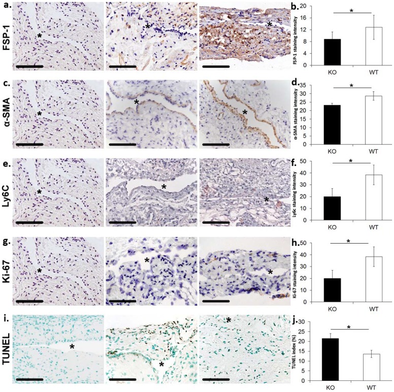
Figure 4. There is a significant decrease in fibroblast, α-SMA, and Ly6C staining accompanied with a decrease in proliferation, and increase in cell death in the outflow vein removed Iex-1 KO mice when compared to WT controls at day 28 after fistula.
a. Representative staining for Fsp-1 from the outflow vein removed from Iex-1 KO (second column) and WT controls (third column) are shown. IgG antibody staining was performed to serve as negative control in the first column. Brown staining cells are positive for Fsp-1. All images are 40X. Scale bar is 100-µms. * indicates lumen. b. Pooled data from the semiquantitative analysis for intensity of Fsp-1 staining in the vessel wall of the outflow vein specimens removed from Iex-1 KO and WT mice. There is a significant decrease in the mean Fsp-1 staining in the Iex-1 KO animals when compared to WT controls (P<0.05). c. Representative staining for α-SMA from the outflow vein removed from Iex-1 KO (second column) and WT controls (third column). IgG antibody staining was performed to serve as negative control in the first column. Brown staining cells are positive for α-SMA. d. Pooled data from the semiquantitative analysis for intensity of α-SMA staining in the vessel wall of the outflow vein specimens removed from Iex-1 KO and WT mice. There is a significant decrease in the mean α-SMA staining in the Iex-1 KO animals when compared to WT controls (P<0.05). e. Representative staining for Ly6C from the outflow vein removed from Iex-1 KO (second column) and WT controls (third column). IgG antibody staining was performed to serve as negative control in the first column. Brown staining cells are positive for Ly6C. f. Pooled data from the semiquantitative analysis for intensity of Ly6C staining in the vessel wall of the outflow vein specimens removed from Iex-1 KO and WT mice. There is a significant decrease in the mean Ly6C staining in the Iex-1 KO animals when compared to WT controls (P<0.05). g. Representative staining for Ki-67 from the outflow vein removed from Iex-1 KO (second column) and WT controls (third column). IgG antibody staining was performed to serve as negative control in the first column. Nuclei staining brown are positive for Ki-67. h. Pooled data for the semiquantitative analysis for intensity of Ki-67 staining in the vessel wall of the outflow vein specimens removed from Iex-1 KO and WT mice. There is a significant decrease in the mean Ki-67 staining in the Iex-1 KO animals when compared to WT controls (P<0.05). i. Representative staining for TUNEL from the outflow vein removed from Iex-1 KO (second column) and WT mice (third column). Negative control is shown where the recombinant terminal deoxynucleotidyl transferase enzyme was omitted in first column. Nuclei staining brown are positive for TUNEL. j. Pooled data from the semiquantitative analysis for intensity of TUNEL staining in the vessel wall of the outflow vein specimens removed from Iex-1 KO and WT mice. There is a significant decrease in the mean TUNEL staining in the Iex-1 KO animals when compared to WT controls (P<0.05). Two-way Student t-test with post hoc Bonferroni's correction was performed. Significant difference from control value is indicated by * P<0.05. Each bar shows mean ± SEM of 4–5 animals per group. For all representative sections * denotes vessel lumen. 40X magnification, scale bar is 100-µms.

