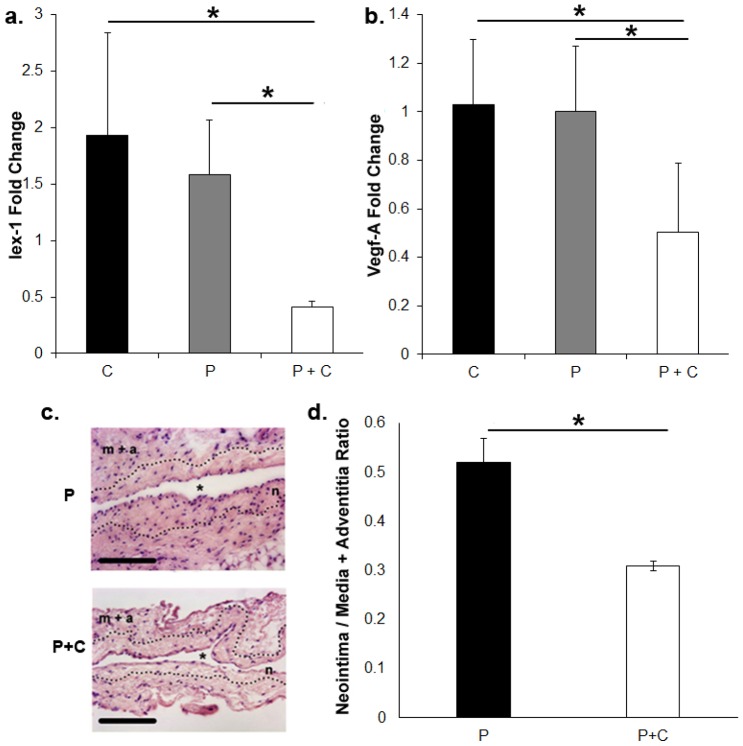
Figure 6. Gene expression of Iex-1 and Vegf-A by qRT-PCR in outflow vein of AVF seven days after adventitial delivery of hydrogel alone, hydrogel with nanoparticle PLGA without calcitriol, or hydrogel with calcitriol in nanoparticle PLGA.
a. Pooled data for Iex-1 expression by qRT-PCR in outflow veins of AVF seven days after adventitial delivery of hydrogel alone (C), hydrogel with nanoparticle PLGA without calcitriol (P), or hydrogel with calcitriol in nanoparticle PLGA (100-µM, P+C). This demonstrates a significant decrease in the mean Iex-1 expression in nanoparticle coated with calcitriol compared to PLGA (P<0.05). b. Pooled data for Vegf-A expression by qRT-PCR in outflow veins treated of AVF seven days after adventitial delivery of hydrogel alone (C), hydrogel with nanoparticle PLGA without calcitriol (P), or hydrogel with calcitriol in nanoparticle PLGA (100-µM, P+C). This demonstrates a significant decrease in the mean Vegf-A gene expression in nanoparticle coated with calcitriol compared to PLGA alone (P<0.05). c. Representative hematoxylin and eosin staining of the outflow vein removed from PLGA (P) and PLGA + Calcitriol (P+C) from 28 days after fistula placement. The neointima (n) is identified from the media and adventitia by the dotted line. m+a is the media and adventitia. * indicates lumen. 40X magnification, scale bar is 100-µms. d. Neointima area/Media + adventitia area ratios for the P and P+C group. There was a significant reduction in the average neointima to media plus adventitia ratio in the calcitriol treated vessels when compared to PLGA alone (P<0.05). Two-way Student t-test with post hoc Bonferroni's correction was performed. Significant difference from control value is indicated by * P<0.05. Each bar shows mean ± SEM of 4-6 animals per group.

