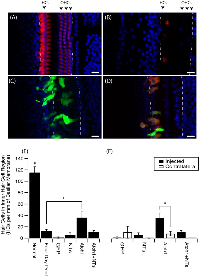
Figure 1. An increase in hair cells in the inner hair cell region after ATOH1 gene therapy.
Example photomicrographs of surface preparations of the basal turn of the cochlea from (A) normal, (B) four day deaf, (C) Ad-GFP treated, (D) Ad-ATOH1 treated animals (red = myosinVIIa, green = GFP, blue = DAPI, dashed lines mark the sensory region, scale bar = 20 µm). (E) When quantified there was a significant loss of IHCs after four days of deafness compared to normal hearing animals. Three weeks post ATOH1-gene therapy there were significantly more myosinVIIa positive cells in the IHC region compared with the four day deaf group (*p<0.05, ANOVA). This number however remained below that observed in normal cochleae which had a greater number of IHCs compared to any other treatment group (#p<0.05, ANOVA). (F) A comparison of treated (injected) versus non-treated (contralateral) cochleae showed ATOH1-injected cochleae to have a significantly greater number of IHCs relative to the contralateral control cochleae (§p<0.05 paired t-test).

