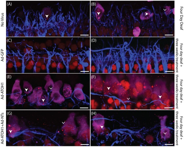
Figure 5. Surface preparations illustrating peripheral fibres in close proximity to HCs with ribbon synapses.
Examples of peripheral fibres (blue) close to myosinVIIa-positive cells (magenta) with CtBP2 puncta (red; filled arrowhead) or without CtBP2 puncta (arrowhead) in the four day deafened (A&B), GFP treated (C&D), ATOH1 treated (E&F) or ATOH1+NTs (G&H) treated cochleae. Scale bar = 10 µm.

