
Figure 6. Resprouting peripheral fibres.
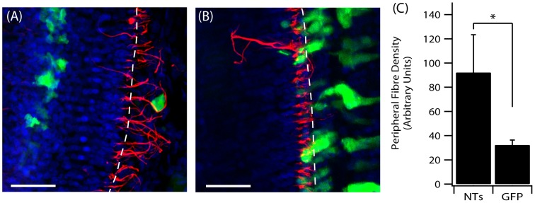
Cochlear surface preparations were stained with anti-NF (red) and DAPI (blue). (A) Peripheral fibre responses to NT-expressing cells in the OC post Ad-NTs treatment. (B) Peripheral fibre response to GFP only expressing cells in the OC post Ad-GFP treatment. The perforated lines indicate the approximate medial edge of the inner pillar cells of the OC. Scale bar = 50 µm. (C) Significantly greater fibre density was observed in close proximity (≤10 µm) to NT-secreting cells compared to GFP-only transduced cells (p<0.05, t-test).