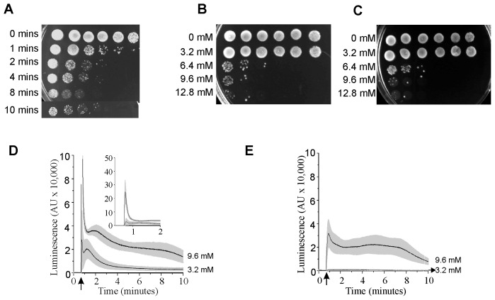
Figure 1. Eugenol toxicity is not dependent on Ca2+ influx.
A) Time dependence of eugenol toxicity. Viability of JK9-3da cells after exposure to 6.4 mM eugenol suspended in Ca buffer for 1, 2, 4, 8 and 10 minutes. Yeast cultures are spotted on to SCM-leu media containing 2% agar; left most spots are growth after 2 days following inoculation with 5 µl of culture. Serial 10-fold dilution of the left most inoculum is shown to the right. B) as A except cells were exposed to varying concentrations of eugenol (as indicated) for 10 minutes in Ca buffer. C) as B except cells were exposed to eugenol in BAPTA buffer. D) Eugenol-induced cytosolic Ca2+ elevation in the presence of extracellular Ca2+ in mid log growth phase yeast cells. Ca2+-dependent aequorin luminescence from Jk9-3da cells in response to 3.2 and 9.6 mM eugenol in Ca buffer. Eugenol was added at 40 seconds (indicated by arrow). Traces represent mean (± SEM) from at least 5 independent experiments. SEM values are illustrated using grey shading. Luminescence was recorded every 0.2 seconds and is expressed in arbitrary units (AU). Inset is data from the main figure on an expanded y axis. E) As D except eugenol was in BAPTA buffer.

