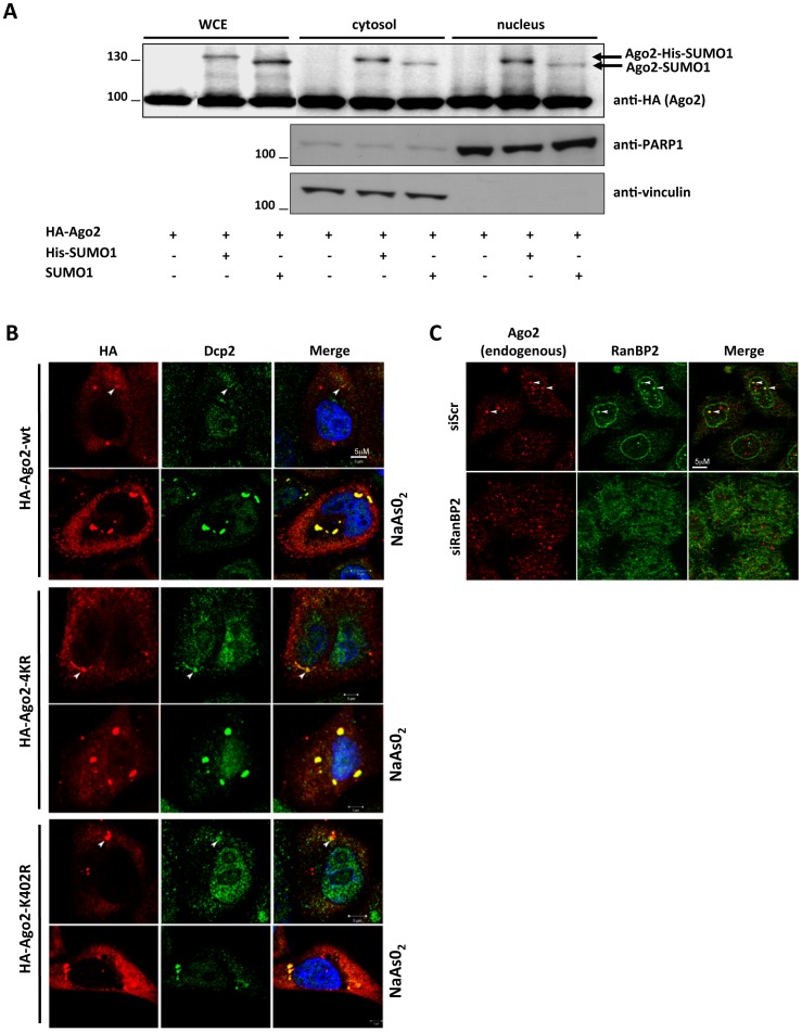
Figure 3. Subcellular localization of SUMO-conjugated Ago2.
(A) Sumoylated Ago2 resides both in the cytosol and nucleus. HeLa cells were transfected with the indicated plasmids and fractionated to analyze nuclear and cytoplasmic proteins. HA-Ago2 was detected by anti-HA antibody. Immunoblots for endogenous PARP1, a nuclear protein, and endogenous vinculin, a cytoplasmic protein, were performed to validate nuclear-cytoplasmic fractionation. (B) Sumoylation is dispensable for subcellular localization of Ago2. HeLa cells were transfected with wild type or sumoylation mutants (4KR and K402R) of HA-Ago2, and treated as indicated (250 µM sodium arsenite -NaAsO2- for 60 min). Ago2 localization was determined by immunofluorescence using the anti-HA antibody (red). The cells were also immunostained for Dcp2 (marking P-bodies in green). The nuclei are stained with DAPI (blue). Ago2 staining was diffuse in the cytoplasm, as well as concentrated in cytoplasmic P-bodies (marked by arrows in untreated cells). NaAsO2 treatment increases size and number of P-bodies. (C) Endogenous Ago2 (red) and RanBP2 (green) colocalize in the nuclei of HeLa cells (indicated by arrowheads).

