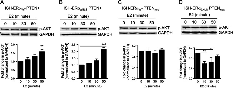
Fig. 4.
E2 stimulation increases phospho-AKT in a PTEN-dependent manner. a ISH-ERαWT PTEN+ and b ISH-ERαΔNLS PTEN+ were starved in serum free media for 24 h followed by E2 treatment for 0, 10, 30, or 50 min. Cells were harvested and analyzed via Western blotting with an antibody specific to phospho-AKT (473) and total AKT. Corresponding graphs below Western blot image represent quantification of fold change ± SD of phospho-AKT normalized to GAPDH (ISH-ERαWT PTEN+ cells, n = 4 (*P < 0.05 by ANOVA) and ISH-ERαΔNLS PTEN+ cells, n = 6 (***P < 0.001 by ANOVA)). c ISH-ERαWT PTENNEG and d ISH-ERαΔNLS PTENNEG cells were starved in serum-free media for 24 h followed by E2 treatment for 0, 10, 30, or 50 min. Cells were harvested and analyzed via Western blotting with an antibody specific to phospho-AKT (473) and total AKT. Corresponding graphs below Western blot image represent quantification of fold change ± SD (n = 3) of phospho-AKT normalized to GAPDH (ISH-ERαWT PTENNEG cells, P = ns by ANOVA and ISH-ERαΔNLS PTENNEG cells, **P < 0.01 by ANOVA). Quantification of fold change ± SD (n = 3) of total AKT normalized to GAPDH resulted in P = ns, by ANOVA, in all cell lines (data not shown)

