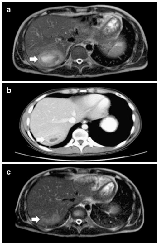
Fig. 1.

a MRI of the abdomen on admission. T2 heterogeneously hyperintense lesion measuring 6.5×5×4.5 cm within hepatic segment VIII, compatible with liver abscess. b CT scan of the abdomen two weeks after removal of percutaneous drain. c MRI of the abdomen one month after discharge. Residual 3.5×1.5 ×1.5 cm area of moderately increased T2 hyperintensity visible at the previous site of the abscess with mild enhancement but no central fluid collection
