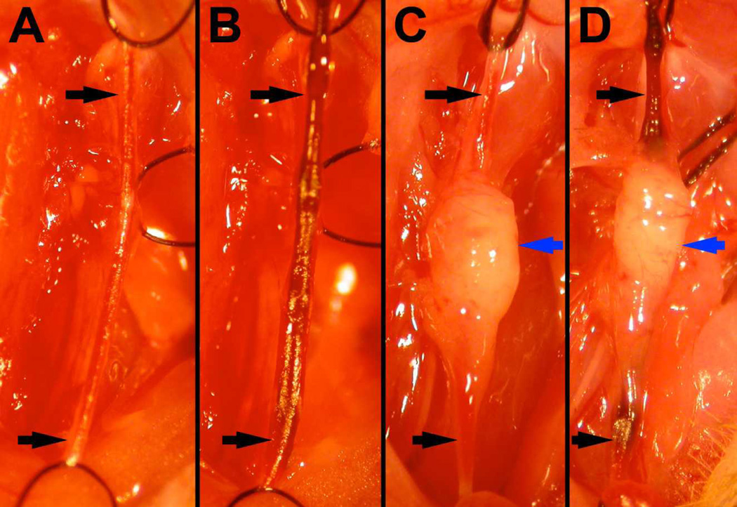
Figure 1.
Surgical images taken during wire injury procedure. Two weeks after either sham transplantation (A, B) or transplantation of 2–3mg of perivascular adipose tissue (C, D), wire injury was performed (B,D). The carotid artery (black arrows) was ligated with silk sutures proximally and distally, relative to the carotid bifurcation. Note that transplanted PVAT is healthy appearing, with incorporated vessels, at the time of wire injury (blue arrows, C & D).

