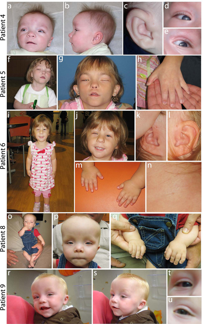
Figure 1.
Photographs of patients documenting features of Peters Plus syndrome. (a–e) Patient 4 with relative macrocephaly, high forehead with sparse eyebrows, hypertelorism, broad nasal bridge, small nose, long, smooth philtrum, thin upper lip (a,b), bilateral preauricular pits (c), and bilateral Peters anomaly with partial clearing of the corneal opacity (d, e); (f–h) Patient 5 with right eye post corneal transplant, left eye enucleated, high forehead, hypertelorism, cupid bow upper lip, long smooth philtrum (f, g) and brachydactyly (h); (i–n) Patient 6 with short stature (i), left ocular prosthesis and right eye post sector iredectomy, microcephaly with micrognathia, a long philtrum (i, j), posteriorly rotated ears with preauricular pits (k, l), brachydactyly (m), and branchial fistula (n) ; (o–q) Patient 8 with short stature (o), broad forehead with sparse eyebrows, relative macrocephaly, a broad nasal bridge, hypertelorism, upslanting palpebral fissues, a simple philtrum with thin lips, low set ears (o,p), and brachydactyly (q); (r–u) Patient 9 with prominent forehead, prominent nose, protruding, thin upper lip, high arched palate, mildly receding chin, small, low set ears (r,s) and Peters anomaly of the right eye only (t,u).

