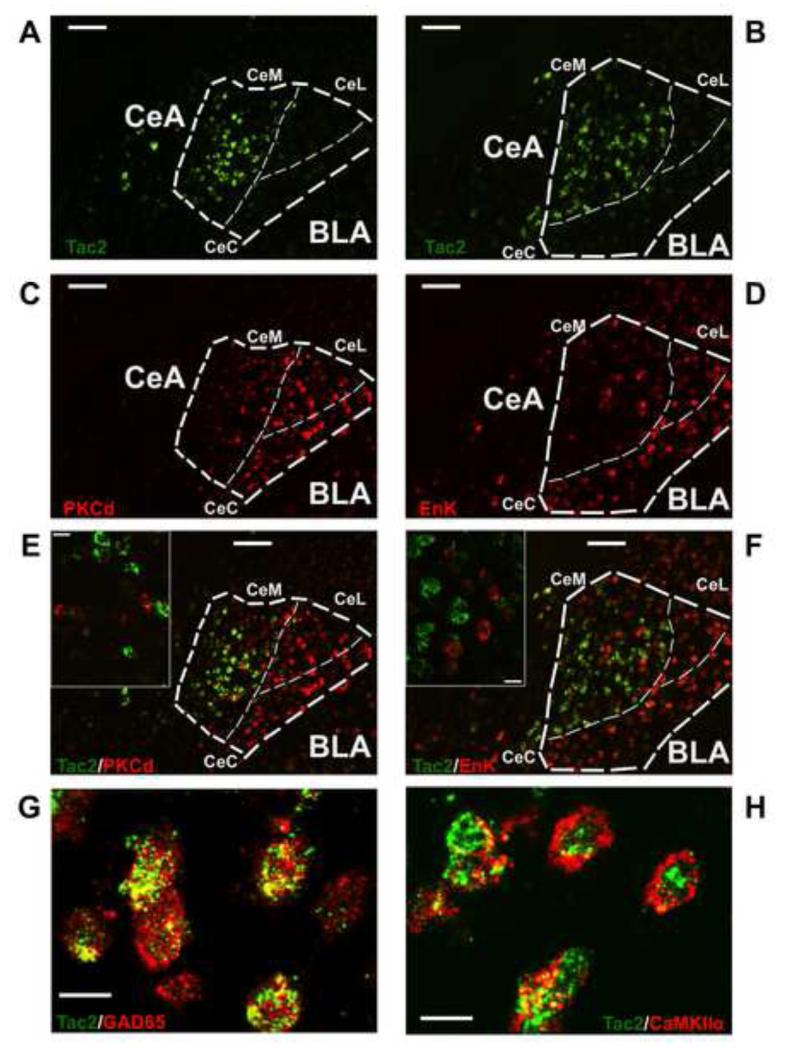
Figure 2. Tac2 is colocalized with glutamate decarboxylase 65 (GAD65) and calmodulin-dependent protein kinase II α (CAMKIIα) but is not colocalized with Protein Kinase C Delta nor Enkephalin-expressing neurons in the CeM.
A and B) Tac2 mRNA expression in the CeA and BLA by non-radioactive fluorescent in situ hybridization (FISH). Scale bar = 100 μm. C) PKCd mRNA expression by FISH. Scale bar = 100 μm. D) Enk mRNA expression by FISH. Scale bar = 100 μm. E) Right, A and C merged showing different pattern of expression of Tac2 and PKCd in the CeA. Scale bar = 100 μm. Left, confocal image showing no colocalization of Tac2 and PKCd in the CeM. Scale bar = 15 μm. F) Right, B and D merged showing different pattern of expression of Tac2 and Enk. Scale bar = 100 μm. Left, confocal image showing no colocalization of Tac2 and Enk in the CeM. Scale bar = 15 μm. G) Confocal image showing colocalization of Tac2 mRNA expression and GAD65 peptide in the CeM. Scale bar = 15 μm. H) Confocal image showing colocalization of Tac2 mRNA expression and CaMKIIα peptide in the CeM. Scale bar = 15 μm. CeM = centro-medial amygdala, CeL = centro-lateral amygdala, CeC = centro-central amygdala, CeA = central amygdala, BLA = basolateral amygdala.

