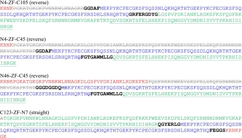
Fig. 2.

Sequences of the designed ZFNs. The ZF proteins are in blue, while NColE7 is divided into three parts: the original N-terminus (in red), the middle part not used in the model (in grey, crossed out) and the original C-terminal part (in green). The linkers are shown with black bold letters. The HNH motif of NColE7 is underlined. The N4–ZF–C105, N4–ZF–C45 and N46–ZF–C45 models are in the reverse orientation, while C123–ZF–N7 is the straight joint of protein sequences. For explanation of the names see the text
