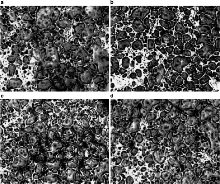
Fig. 1.
TRAP staining of bone marrow macrophages. Bone marrow macrophages were collected and spun at 1300 rpm for 8 min at room temperature, 660,000 cells were seeded in each well of the six-well plate with M-CSF (20 ng/ml) and RANKL (100 ng/ml). Shown is a representative picture of TRAP staining repeated at least three different times, a TRAP staining after 6 days of differentiation; b TRAP staining of osteoclasts incubated for the last 3 days with 100 µM tryptophan. c TRAP staining of osteoclasts incubated for the last 3 days with 100 µM phenylalanine. d TRAP staining of osteoclasts incubated for the last 3 days with 100 µM Tyrosine. Photomicrographs showed multinucleated osteoclastic cells in the control and the treated groups and of note; no changes were detected in the morphology of the cells

