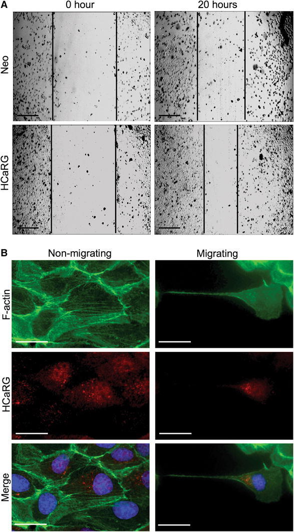
Fig. 3.

HCaRG stimulates renal cell migration. a Wound healing assay in Neo- or HCaRG-plasmid-transfected HEK-293 cells. HCaRG markedly accelerated wound healing after 20 h compared to Neo-controls. Scale bars 50 μm. b Intracellular location of F-actin (green) and HCaRG (red) in migrating MDCK cells during the wound-healing process. In non-migrating cells, HCaRG is located in the perinuclear space, and organized actin fibers are clearly visible. In the migrating cell, intracellular actin fibers are de-polymerized and short F-actin filaments are observed in the perinuclear space. HCaRG localizes with short F-actin filaments between the nucleus and elongated tip. Nuclei are stained by DAPI (blue). Scale bars 25 μm (color figure online)
