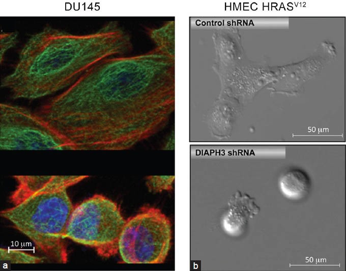
Figure 1.

(a) Mesenchymal (top) and amoeboid (bottom) subpopulations occur naturally within DU145 PCa cells. Cells were stained with antitubulin (green) and phalloidin (red) to demonstrate differences in tubulin and actin cytoskeletal organization. The cell nucleus is blue (DAPI). Note the difference in size between mesenchymal and amoeboid cells. (b) Mesenchymal (top)-amoeboid (bottom) transition in HMEC-HRasV12-transformed HMECs upon DIAPH3 silencing.23 DAPI: 4’,6-diamidino-2-phenylindole; HMEC: human mammary epithelial cells; PCa: prostate cancer.
