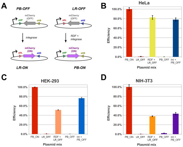
Figure 2. Plasmid inversion assays.
(A) Simplified diagram of detection schemes for PxB and LxR inversion. In the un-flipped state, mCherry is not expressed, as its ORF is inverted relative to the upstream promoter. When acted upon by Int or RDF:Int (for PB-OFF and LR-OFF, respectively), the mCherry ORF is flipped, which leads to its expression, detectable via fluorescence. Assays with these plasmids were conducted in HeLa (B), HEK-293 (C) and NIH-3T3 (D) cells. The percentage of cells expressing mCherry was measured in triplicate for each plasmid combination and then normalized to the positive control, the PB-ON transfection efficiency of same cell type. Representative data from one of three independent trials is shown here. ‘RDF’ and ‘Int’ refer to the pCS-kRI and pCS-kI plasmids, respectively. The error bars indicate the standard error of the mean.

