
Figure 6.
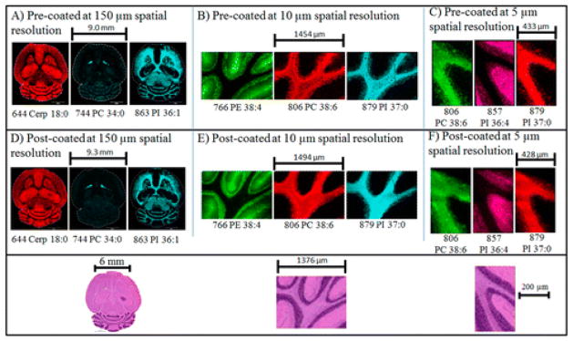
Comparison of imaging results of mouse brain serial sections with matrix pre- and post-coated targets at different spatial resolutions. Ion images were collected in negative ion mode. Tentative identification was based on previous MS/MS analyses of lipids at these masses in mouse brain. Below: H&E optical scanned images from a serial section are shown at the bottom of the figure.