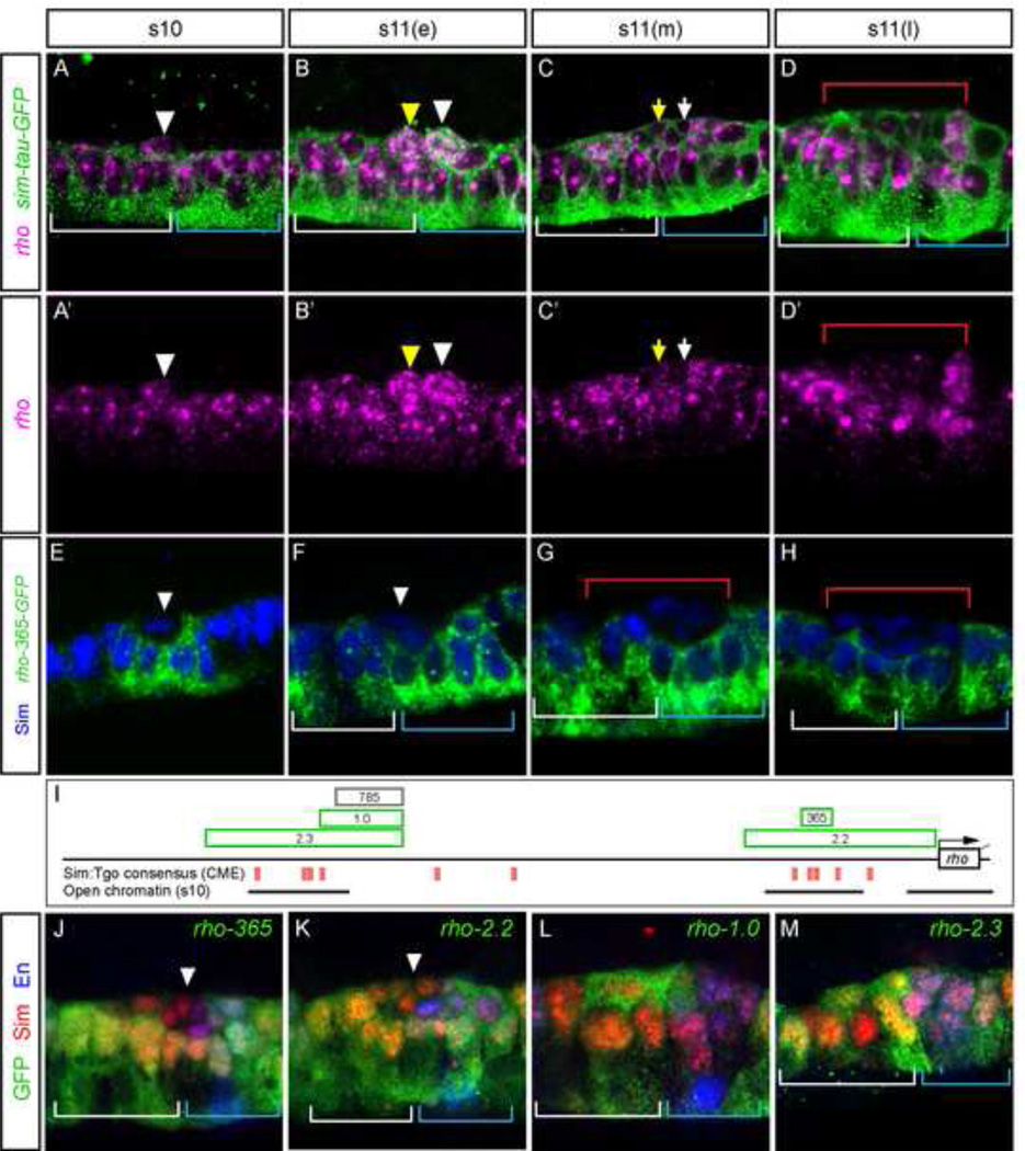
Figure 4. Multiple enhancers collaborate to control rhomboid midline primordium expression.
(A-D’) Expression dynamics of rho (magenta) is shown in representative segments in sim-Gal4 UAS-tau-GFP (sim-tau-GFP) embryos also stained with anti-tau (green). Stages shown are: (A,A’) stage 10, (B,B’) early stage 11 showing a burst of rho expression in MP3 (yellow arrowhead) and MP4 (white arrowhead), (C,C) mid stage 11 in which neuronal progeny (arrows) show reductions in expression, and (D,D’) late-stage 11 showing low levels of rho expression in neurons (below red bracket). (E–H) rho-365-GFP embryos stained for GFP RNA (green) and Sim protein (blue). (E) Arrowhead indicates the presence of strong GFP in a small number of midline cells at stage 10. (F) GFP expression expands posteriorly in midline cells, but is restricted from iVUM4/mVUM4 (white arrowhead). (G,H) Reduction of GFP expression is apparent in midline neurons beneath the red bracket, but is maintained in AMG (white bracket) and PMG (blue bracket). (I) Schematic of rho locus showing features related to gene expression. Genomic fragments with midline activity are indicated as green boxes; rho-785 does not have midline activity. Consensus Sim:Tgo binding site matches (ACGTG) are indicated as red boxes, and the presence and extent of open chromatin regions (DNase I hypersensitive sites, 5% FDR, adapted from genome.ucsc.edu) at stage 10 are indicated by lines. (J–M) Embryos containing rho midline fragments driving GFP expression at mid-stage 11 stained for GFP, Sim, and En. (J,K) Arrowheads point to neurons with reduced expression.

