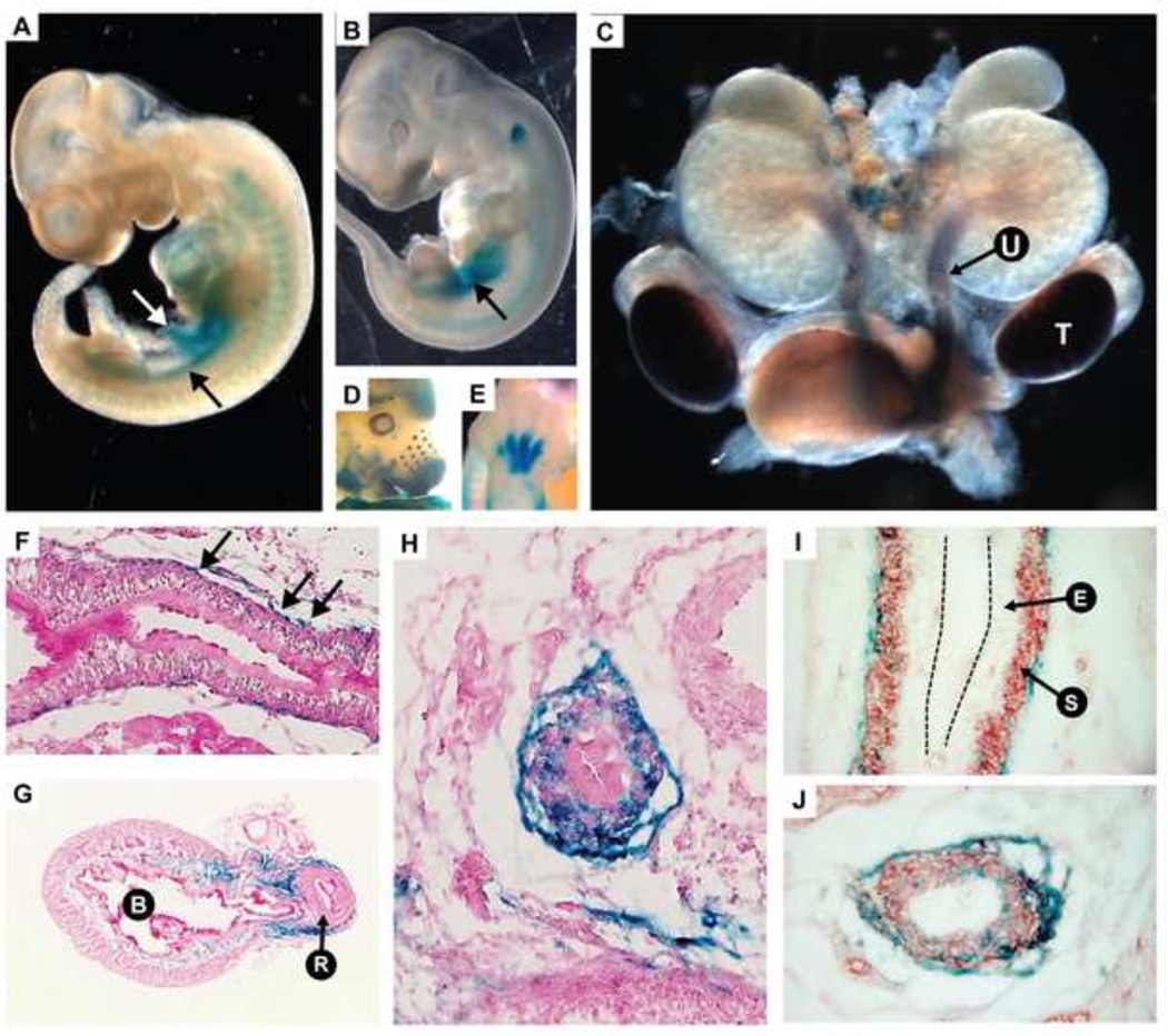
Fig. 5.
The ECR1 enhancer which spans the 12Gso breakpoint contains a urogenital-active enhancer element. (A) At embryonic day E10.5, the enhancer directs LacZ expression in the urogenital ridge (black arrow) and the vitelline vein (white arrow). (B) Expression at E11.5 is primarily localized to the urogenital mesenchyme (black arrow). (C) E14.5 dissected urogenital system shows strong staining in the ureter mesenchyme “U” and testes “T”. (D–E) LacZ is also expressed in the developing whisker buds, maxilla and mandible, eyecup, forebrain, and limbs as previously reported for the Tbx18 mRNA (Airik et al., 2006; Kraus et al., 2001). (F–H) Sections of LacZ stained E16.5 ureters and bladder showing expression in the ureter mesenchyme. (F) Black arrows indicating β-galactosidase stained cells along the length of the ureter. (G) Bladder indicated “B” and urethra is indicated by “R”. Blue staining is observed in a portion of the bladder stroma. (I) The urinary epithelium is labeled “E” and the differentiated smooth muscle layer is labeled “S”. The black dotted lines indicate the apical surface the urinary epithelium. (I–J) E16.5 ureters co-stained with LacZ (blue) and anti-SMA (red). (J) In the ureter section closest to the bladder, LacZ positive cells are dispersed through the mesenchyme.

