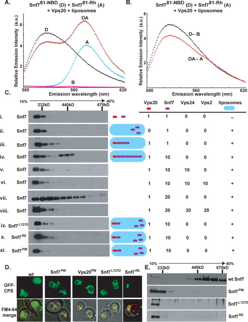
Fig. 5. Oligomerization of Snf7.

(A) Emission spectrum (λex = 478 nm) of the DA sample (red) containing 320 nM Snf781-NBD, 4.2 μM of Snf781-Rh, 0.5 μM Vps20, and 1.5 mM PC/PS/PI. Unlabeled Snf781-C was substituted for one or both of the fluorescent derivatives to form the D (black), A (cyan), and B (magenta) samples. (B) The normalized net D (black) and net DA (red) emission spectra obtained from the samples in A. (C) Purified ESCRT-III proteins were mixed in different combinations and using different molar ratios (as indicated) in the presence or absence of PC/PS/PI. The liposomal fraction was isolated by centrifugation, detergent-solubilized, and analyzed by velocity sedimentation. (D) In wild-type (wt) cells, GFP-tagged carboxypeptidase-S (GFP-CPS) accumulates in the vacuolar lumen, while the FM4-64 dye stains the limiting membrane of the vacuole. In snf7Δ cells expressing the different mutant forms of Snf7 or in vps20Δ cells expressing the Vps20PW mutant, GFP-CPS co-localizes either with the limiting membrane of the vacuole or the class E compartment. (E) Endosome (P13) fractions prepared from vps4Δsnf7Δ cells expressing either wt Snf7 (top panel) or the different Snf7 mutants were detergent solubilized, and analyzed using glycerol velocity gradients.
