
Fig. 6. ESCRT-III assembly, disassembly and liposome deformation.
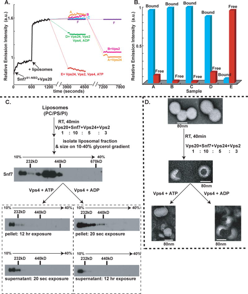
(A) Time- and component-dependent emission intensity profiles. Each reaction sample (A–F) contained 540 nM Snf781-NBD mixed with Vps20 at time 0. At 600 sec, 1.5mM PC/PS/PI was added to each sample. At 1200 sec, additions were made as follows: sample A (orange trace) to 180 nM Vps2, 1 μM Vps4, 1mM ATP; B (magenta) to 270 nM Vps24, 1 μM Vps4, 1mM ATP; C (cyan) to 270 nM Vps24, 180 nM Vps2, 1 μM Vps4 (E233Q), 1mM ATP; D (green) to 270 nM Vps24, 180 nM Vps2, 1 μM Vps4, 1mM ADP; E (red) to 270 nM Vps24, 180 nM Vps2, 1 μM Vps4, 1mM ATP; and F (purple) received only buffer. After incubation (37°C, 45 min), emission was re-measured from 3900–4500 sec at 22°C. Samples A, B, and F then received 270 nM Vps24, 180 nM Vps2, or buffer, respectively, were incubated (37°C, 45 min), and emission was re-measured from 7200–7800 sec. (B) In some experiments, samples were analyzed by Sepharose CL-2B gel filtration chromatography after completing the first 45 min incubation. Protein was detected by NBD emission intensity, and liposomes by light scattering (cyan). The red and blue bars indicate the relative amounts of free Snf781-NBD and liposome-bound Snf781-NBD respectively. (C) Outline of the in vitro ESCRT-III disassembly assay. ESCRT-III complex was assembled on liposomes using purified ESCRT-III proteins and centrifuged to separate the liposome-bound complex The liposome-bound ESCRT-III complex was treated with either (i) Vps4 and ATP or (ii) Vps4 and ADP for 45 min at 37°C. Following incubation, the samples were centrifuged to separate the liposome-bound proteins (“pellet”) from free ESCRT-III proteins (“supernatant”), and then further analyzed by velocity sedimentation. (D) Negative stain EM analyses of PC/PS/PI liposomes, ESCRT-III bound liposomes, ESCRT-III-bound liposomes treated with 1μM Vps4 + 1mM ATP, and ESCRT-III-bound liposomes treated with 1 μM Vps4 + 1mM ADP.