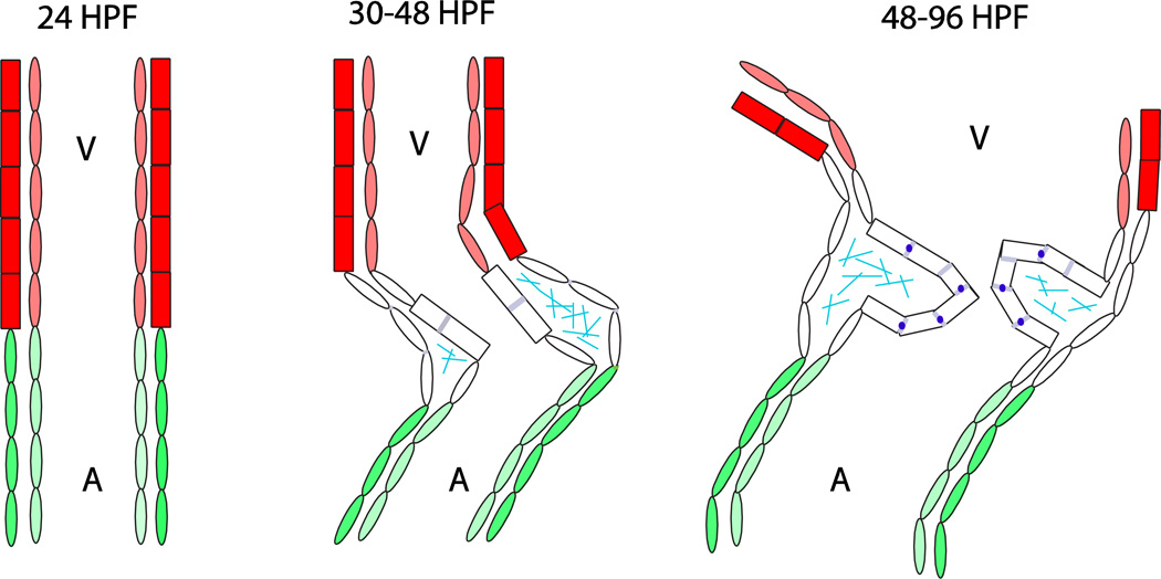
Figure 1.
Morphological changes in the zebrafish atrioventricular canal between 24 and 96 hours post-fertilization. Dark red = ventricular myocardium; light red = ventricular endocardium; green = atrial myocardium; light green = atrial endocardium; rectangles = cuboidal cells; ovals = squamous or trapezoidal cells; grayish blue = Dm grasp expression; sky blue = chondroitin sulfate expression; dark blue = Z01 expression; A = atrium; V = ventricle.

