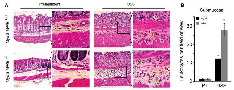
Fig. 2.
DSS causes a greater influx of leukocytes into the colonic submucosa of Myc 3′ WRE−/− mice in comparison with WT mice. a H&E-stained sections of colonic tissue prepared before treatment and after five days of DSS treatment. Each panel is a representative image from n = 3 mice examined per genotype and enlargements of the boxed regions are placed to the right of each image. Yellow arrows identify representative leukocytes in each image. b Quantification of leukocytes in 10 fields of the colonic submucosa. Error is standard error of the mean (*p < 0.0005)

