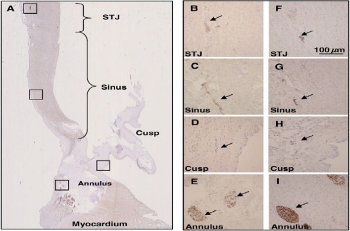
Figure 7.
Presence of sympathetic nerves in the aortic root. Panel A shows the whole aortic root (stained for tyrosine hydroxylase), Panels B–E (stained for tyrosine hydroxylase) and F–I (stained for neurofilament protein) show nerve bundles at a higher magnification in the sinotubular junction (STJ) sinus area, hinge region of the cusp, and in the annular region (from Chester et al., 2008). 118

