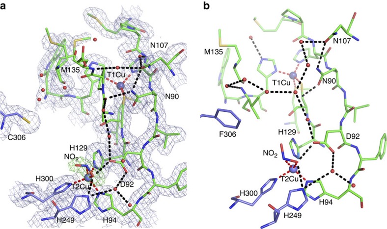
Figure 2. Comparison of the nitrite-binding mode in as isolated F306C mutant and wild-type AxNiR.
2Fo–Fc electron density maps contoured at 1σ level for both (a) as-isolated F306C mutant and (b) wild-type AxNiR. The Cu sites are shown as blue spheres with coordination bonds shown as dashed red lines. Important hydrogen bonds are shown as dashed black lines. 1Fo–Fc omit map for nitrite is shown in green at 4σ level. Note that crystals of F306C were grown in the presence of PEG that prevents nitrite diffusion into crystals of wild-type AxNiR10, consistent with the mutation resulting in enhanced access to the active site.

