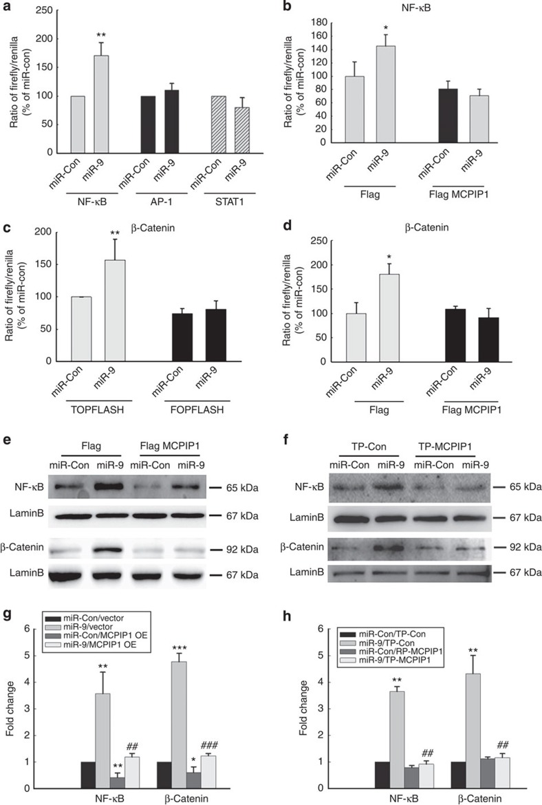
Figure 4. MiR-9-mediated activation of NF-κB and β-catenin involves suppression of MCPIP1.
(a) BV-2 cells transfected with NF-κB-TK-Luc, AP-1-TK-Luc or GAS-TK-Luc constructs were cotransfected with miR-9 for 24 h followed by reporter gene activity analysis using Luciferase assay. (b) MCPIP1 Δ3′UTR overexpression inhibited NF-κB activation. (c) BV-2 cells transfected with Topflash or Fopflash constructs were co-transfected with miR-9 for 24 h followed by reporter gene activity analysis using Luciferase assay. (d) MCPIP1 Δ3′UTR overexpression inhibited activation β-catenin. All the data are indicated as mean±s.d. of four individual experiments. *P<0.05, **P<0.01 versus microglial transfected with miR-control using Student’s t-test. (e) Transfection of microglia with MCPIP1 Δ3′UTR construct resulted in inhibition of miR-9 mediated nuclear translocation of NF-κB and β-catenin. (f) Transfection of microglia with TP-MCPIP1 oligo resulted in inhibition of miR-9-mediated nuclear translocation of NF-κB and β-catenin. (g and h) Densitometric analysis of NFκB and beta catenin from four separate experiments. All the data are indicated as mean±s.d. of four individual experiments. *P<0.05, **P<0.01, ***P<0.001 versus microglial transfected with miR-control and vector group; ##P<0.01, ###P<0.001 versus microglial transfected with miR-9 and vector group using one-way analysis of variance followed by Holm–Sidak test.

