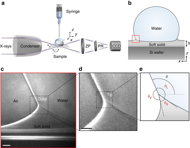
Figure 1. High-resolution X-ray imaging for wetting ridge formation.
(a,b) Schematic illustrations of (a) transmission X-ray microscopy (TXM) and (b) a sessile drop on a soft substrate. TXM consists of a capillary condenser, a motorized sample stage, a zone plate (ZP), a phase ring (PR) and a CCD camera. A wetting ridge (red square in b) is formed by the surface tension of a water drop at the contact line. (c,d) Demonstration of a directly visualized (c) wetting ridge with (d) a bent cusp (magnified image of the dashed square in c) and an asymmetric tip (dashed square in d). (e) Extraction of three interfaces from the ridge tip (magnified image of the dashed square in d), which enables us to measure the macroscopic (θ) and the microscopic (θS, θV and θL) contact angles. (c,d) Scale bars, 2 and 1 μm, respectively.

