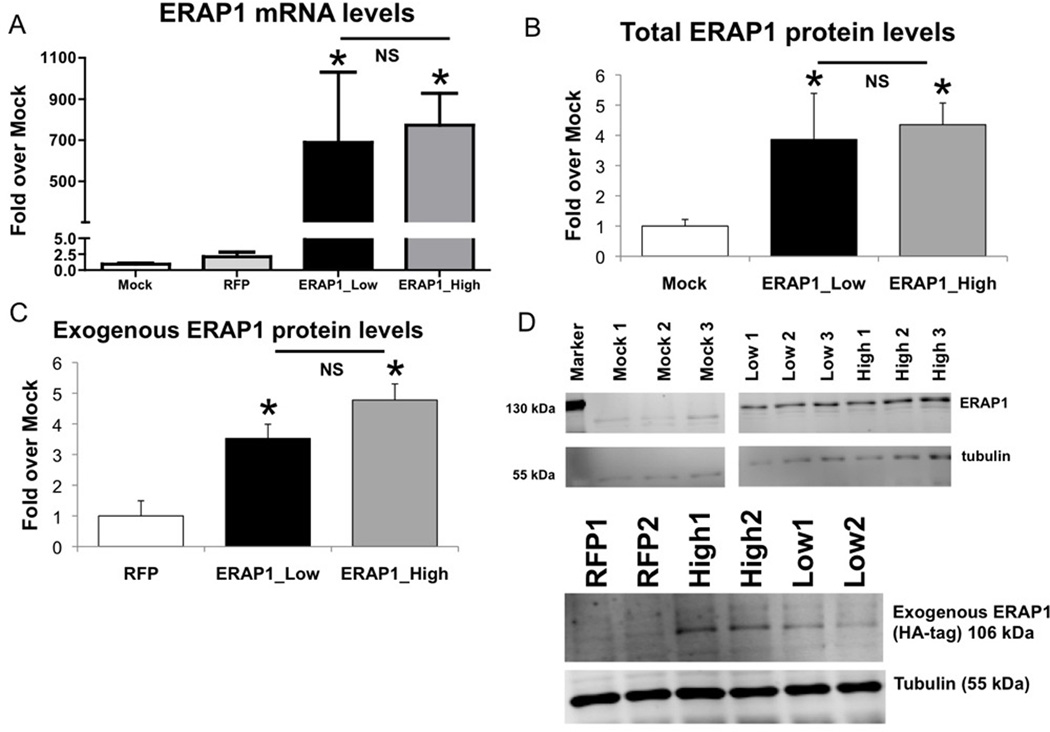
Figure 2. Transfection of HeLa-Kb-B27/47 cells with ERAP1 alleles results in significant increases of ERAP1 mRNA and protein levels, increases which are not different between ERAP1 alleles.
HeLa-Kb-B27/47 cells were transiently transfected with plasmids expressing ERAP1_Low or ERAP1_High alleles (or no ERAP1). RNA and protein samples were harvested at 24 hours post transfection as described in Materials and Methods. Statistical analysis was completed using two-tailed Student’s t-test, * - represent significant difference in ERAP1 levels as compared to control (mock or empty pTracer-RFP transfection), p<0.05; n=2 independent experiments performed. (A) Levels of ERAP1 mRNA are shown for each transfection group. About 700-fold induction in ERAP1 mRNA levels were noted for ERAP1_Low and ERAP1_High transfected cells, as compared to mock transfected cells. (B) Total ERAP1 levels were measured by Western blot. Significant ~3–4 fold induction in total ERAP1 protein levels were noted for ERAP1_Low and ERAP1_High transfected cells, as compared to mock transfected cells. (C) Exogenous ERAP1 levels were measured by Western blot with anti-HA-tag antibody. Significant exogenous ERAP1 levels were found in cells transfected with ERAP1_Low and ERAP1_High. (D) Representative Western blots for total ERAP1 (top) and exogenous ERAP1 (bottom) are shown. ERAP1 bands (106 kDa) and tubulin bands (55 kDa) are depicted.

