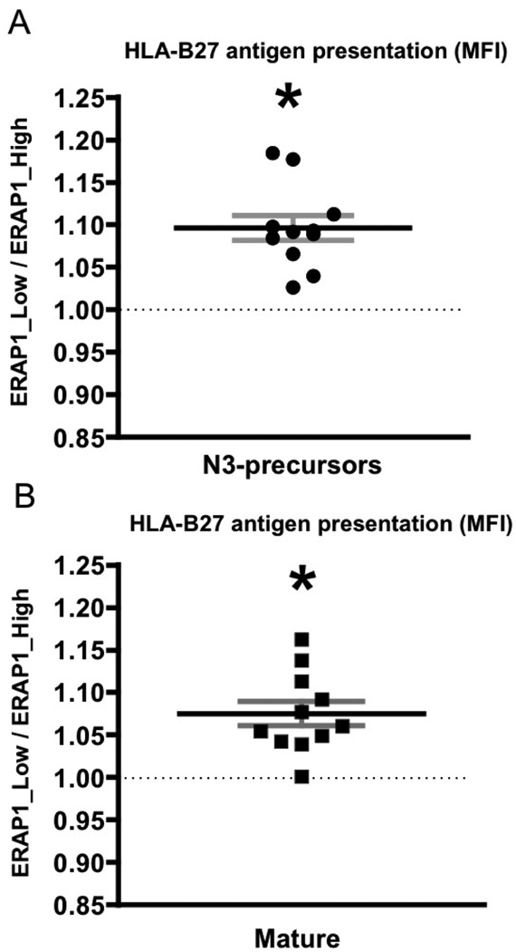
Figure 3. ERAP1_High reduces HLA-B27 antigen presentation in intact cells as compared to ERAP1_Low for vast majority of HLA-B27-restricted peptides.
HeLa-Kb-B27/47 cells were transiently transfected with plasmids expressing HLA-B27-specific peptides (with ER-signal sequence) with GFP and either empty vector (control) or the indicated ERAP1 allele with RFP. At 48 hours post transfection surface HLA-B27 surface expression on double-transfected (GFP+RFP+) was analyzed by flow cytometry using the ME-1 antibody. Peptides were transfected either as (A) N3-extended precursors or (B) “mature” epitopes. Ratio in mean fluorescent intensity (MFI) between ERAP1_Low and ERAP1_High variants is depicted. Combined graph is shown (n=11). Bars represent mean ± SEM. Statistical analysis was completed using one sample t-test [(mean ratio – 1)/SE)]; * - represent significant differences in HLA-B27 surface expression mediated by ERAP1 polymorphism (i.e. fold ratio ERAP1_Low/ERAP1_High is significantly different from 1, dashed line), p<0.05.

