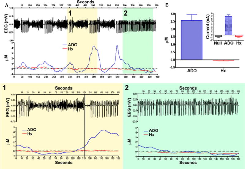
Figure 4. Biosensor amperometric separation of adenosine and hypoxanthene (Hx) during epileptiform events.

(A) Continuous fixed potential amperometry (FPA) recording over 900 seconds demonstrating 5 epileptiform events in which (1) represents a higher magnification of such an event, and (2) shows continuous spiking at the end. Note there is little variation in Hx, but adenosine consistently varies with the epileptiform activity; (B) adenosine and Hx micromolar change with event termination, 95% CI for adenosine is 1.7 to 3.4 μM and for Hx is −0.2 to 0.1. Inset shows variation in raw current (nA) of the FPA recordings. Note that there is a general decrement with null and Hx as is typical with amperometry.
