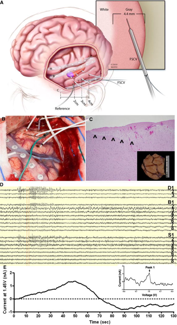
Figure 5. Human Intraoperative Fast Scan Cyclic Voltammetry (FSCV) Recording.

Representative intraoperative human FSCV recorded by the wireless instantaneous neurochemical concentration sensing system (WINCS): (A) a schematic demonstrating the human intraoperative implantation, experimental setup, illustrating the implantation depth electrophysiology electrodes and glass-insulated CFM in the gray matter of temporal cortex targeted for resection for seizure control; (B) intraoperative photo of human surgery utilizing FSCV. A glass capillary carbon fiber microelectrode (*green wire) is delivered into the lateral neocortex near the electrocorticography electrode S3 during pre-lobectomy electrocorticography. FSCV is performed relative to a stainless steel electrode. There is an eight contact strip over the inferior temporal gyrus (right of exposure), an eight contact strip over the superior temporal gyrus (most visible in center of exposure) and a separate strip is over the interior frontal lobe. There are 3 mesial temporal depths in the exposure (white leads) penetrating the middle temporal gyrus; (C) 10x hematoxylin and eosin stain of electrode insertion site on pathology. Note the entire FSCV electrode tract is within grey matter (inset shows the gross anterior temporal lobe specimen with methylene blue at the insertion site); (D) 140 seconds of electrocorticography demonstrating spontaneous epileptiform activity with that from 7 to 30 seconds most prominent in the mesial temporal lobe (D1–3) with associated lateral neocortical activity (S1–3). (E) Current (at 1.45 V or Peak 1 of adenosine) vs. time plot corresponding in time to the above (D) EEG. Note there appears to be an increase in Peak 1 of adenosine with the peak near the termination of this epileptiform event in patient 2. Inset: unfolded voltammogram of the signal at 53 seconds, when scanning from −0.4 to 1.5, showing a unique oxidation peak at 1.45 Volts.
