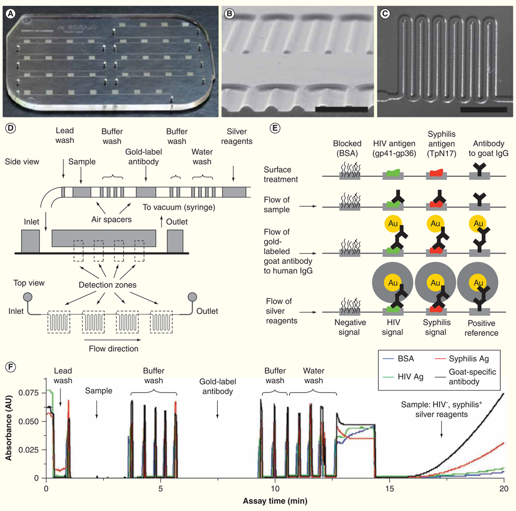
Figure 2. Integrated microfluidic system for multiplexed detection of HIV and syphilis.
(A) Photograph of microfluidic chip. (B) Cross-section of microchannels. Scale bar, 500 µm. (C) The design of channel meanders. Scale bar, 1 mm. (D) Schematic diagram of passive fluid delivery of preloaded reagents over four detection zones based on vacuum generated by a disposable syringe. (E) Illustration of reactions at different detection steps. Signal amplification was achieved by the reduction of silver ions on gold nanoparticle-conjugated antibodies. Signals can be read quantitatively with low-cost optics or qualitatively by eyes. (F) Real-time monitoring of absorbance signals at the detection zones.
Adapted with permission from [63] © Macmillan Publishers Ltd. (2011).

