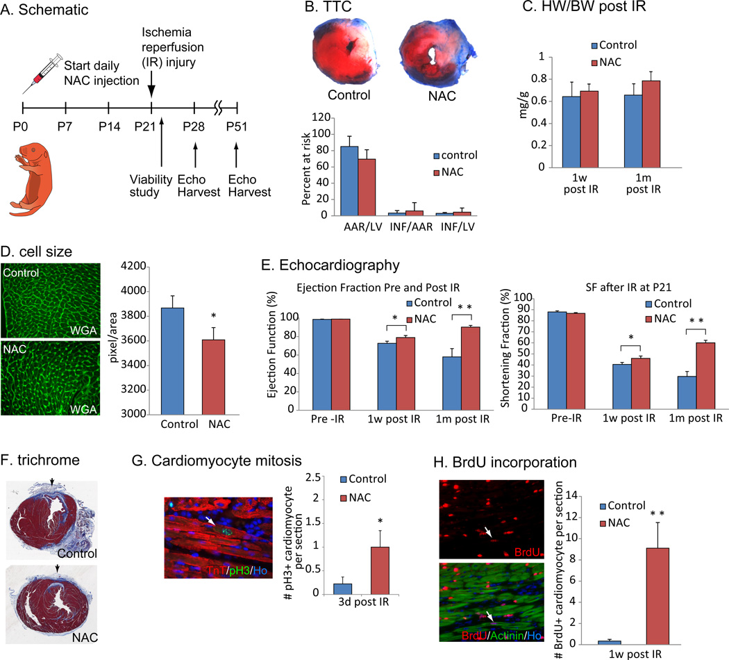
Figure 6.
Scavenging ROS extends postnatal cardiac regeneration window. (A) Animals were treated with NAC from P0 and subjected to ischemia reperfusion (IR) surgery at P21. Viability was investigated 24 hours after injury to evaluate the consistency of the IR surgery, and then cardiac function was examined by echocardiography at 1 week and 1 month after injury. (B) TTC staining showed no significant difference in the size of injury between control and NAC-pretreated hearts. (C) HW/BW ratio at 1 week or 1 month after IR surgery between control and NAC-pretreated mice was not changed. (D) WGA staining showed a decrease in cell size in NAC treated hearts 1 month after IR injury compared to control hearts, indicating the NAC treatment did not induce cardiomyocyte hypertrophy. (E) Left ventricular systolic function quantified by ejection fraction (EF) and shortening fraction (SF) (n=6 per group) before IR surgery (left), 1 week after surgery (middle) and 1 month after surgery (right) showed more functional recovery in NAC-pretreated hearts compared with control hearts. (F) Histological analysis with Masson trichrome staining showed reduced fibrotic scar formation in NAC treated hearts. (G) Cardiomyocyte mitosis was increased in NAC-treated mice as indicated by anti-pH3 and anti-TnT co-immunostaining at 3 days after IR injury. (H) Immunostaining with anti-BrdU and anti-Actinin antibodies showed increased cardiomyocyte BrdU incorporation NAC-pretreated heart. Error bars indicate SEM. *p < 0.05; **p < 0.01.

