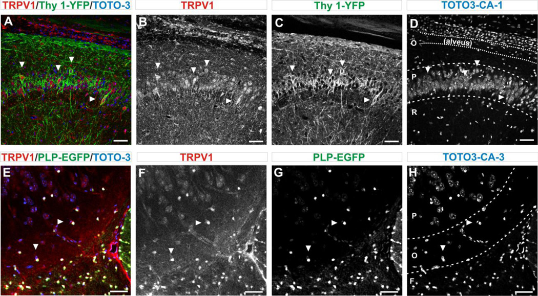
FIGURE 8. OVERALL EXPRESSION OF TRPV1 CHANNELS IN NEURONS AND OLIGODENDROCYTES (ODC).
A. Immuno-labelling for TRPV1 in CA1 pyramidal cell layer of the hippocampus in the Thy1-YFP transgenic mouse show co-expression of neural marker Thy-1-YFP (green) and TRPV1 channels (red). Arrows indicate examples of cells in which co-expression is clear. Sub-regions of hippocampus CA1 are demarcated in D. Confocal images are 25X magnification/ 0.7X field. Scale bar = 50 µm.
B. Immuno-labelling for TRPV1 channels with anti-TRPV1 antibody show expression in the pyramidal layer. Arrows indicate examples of cells in which expression is clear. Scale bar = 50 µm.
C. The Thy1-YFP tracer showing positive neurons along the pyramidal cell layer. Scale bar = 50 µm.
D. Staining with the nuclear marker TOTO3 show the pattern of cell distribution. The organization of the hippocampus was demarcated: (O) stratum oriens containing the alveus, (P) stratum pyramidal and (R) stratum radiatum. Arrows indicate nuclei from cells that co-express Thy1-YFP and TRPV1 markers. Scale bar = 50 µm.
E. Co-expression of TRPV1 channels (red) and the ODC tracer PLP-EGFP (green) in the fornix and CA3 region, using a transgenic reporter mouse (Mallon et al., 2002). Arrows indicate examples of cells in which co-expression is clear. Sub-regions of the hippocampus CA3 are delineated in H. Confocal images are 25X magnification/ 0.7X field. Scale bar = 50 µm.
F. Immuno-labeled for TRPV1 channels with anti-TRPV1 antibody show greater channel expression in small round ODC and a large blood vessel. Arrows indicate examples of cells in which expression is clear.
G. Typical pattern of expression of ODC with greater number of ODC in the fornix and with few ODC that can be seen in both the stratum oriens and pyramidale. Arrows indicate examples of cells in which expression is clear.
H. The nuclear marker (TOTO3) for corresponding area of the hippocampus. CA3 structural elements were demarcated: (F) fornix, (O) stratum oriens and (P) stratum pyramidale. Arrows indicate examples of nuclei from cells in which co-expression is indicated in E, F and G.

