
Fig.3. Wee1B-dependent inactivation of MPF during egg activation.
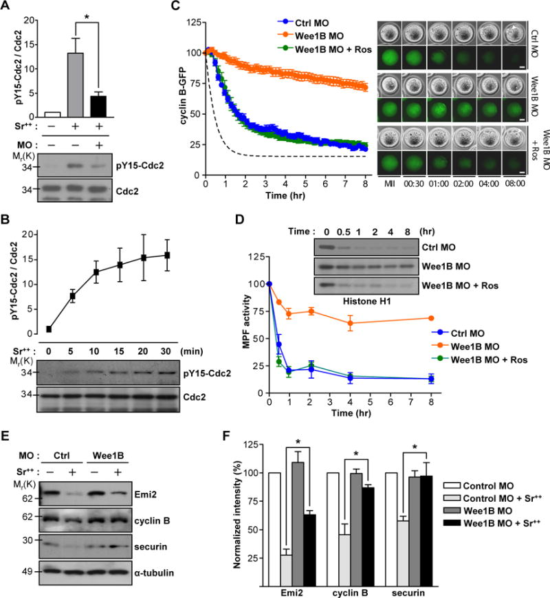
(A) Non-injected or Wee1B MO injected MII oocytes were activated with strontium for 20 min and subjected to SDS-PAGE followed by immunoblotting for Cdc2 and tyrosine 15 phosphorylation (pY15) of Cdc2. (B) MII oocytes activated with strontium were prepared at different time after activation and subjected to SDS-PAGE followed by immunoblotting for Cdc2 and pY15 Cdc2. Data are the mean ± SEM from three independent experiments. A representative image is shown. (C and D) GV oocytes injected with Wee1B MO and cyclin B-GFP mRNA were in vitro matured and activated with strontium at time t=0. (C) GFP levels were measured every 10 min for 8 hours. Average of MPF activity in control oocytes from (D) is shown with dotted line. Data are mean ± SEM of ten oocytes from two independent experiments. Representative images are shown in right panel. Pronucleus is indicated by arrowheads. Bar, 20μm. (D) Oocyte lysates were incubated with [γ-32P]ATP and either Histone H1 or myelin basic protein (MBP) (see fig.S4). Phosphorylation was detected by SDS-PAGE and autoradiography. Representative images from two independent experiments are shown. (E) In vitro matured MII oocytes injected with control or Wee1B MO at GV stage were incubated with (+) or without (−) strontium for 2 hours. Emi2, cyclin B and securin levels were determined by immunoblots. Each lane contains 100 oocytes. Representative images from three independent experiments are shown. (F) Band intensities of Emi2, cyclin B and securin were quantified and normalized to controls. Data are the mean ± SEM from three independent experiments. *p<0.05.