
Figure 1.
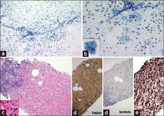
(a and b) Fine-needle aspiration of the mass demonstrate monomorphic lymphocyte population admixed with scattered histiocytes in a clear background. Inset: Histiocyte with refractile cytoplasmic crystals (Diff Quik, ×100 and ×200). (c) Core biopsy showing monomorphic lymphoid aggregate admixed with sheets of histiocytes (H and E, ×100). (d) Immunohistochemistry stains on the core biopsy showing kappa light chain restriction in the lymphocytes as well as in cytoplasmic crystals within histiocytes (kappa and lambda light chain, ×100). (e) CD68 stain performed on the core biopsy highlights CD68+ histiocytes (CD68, ×100)