Fig. 4.
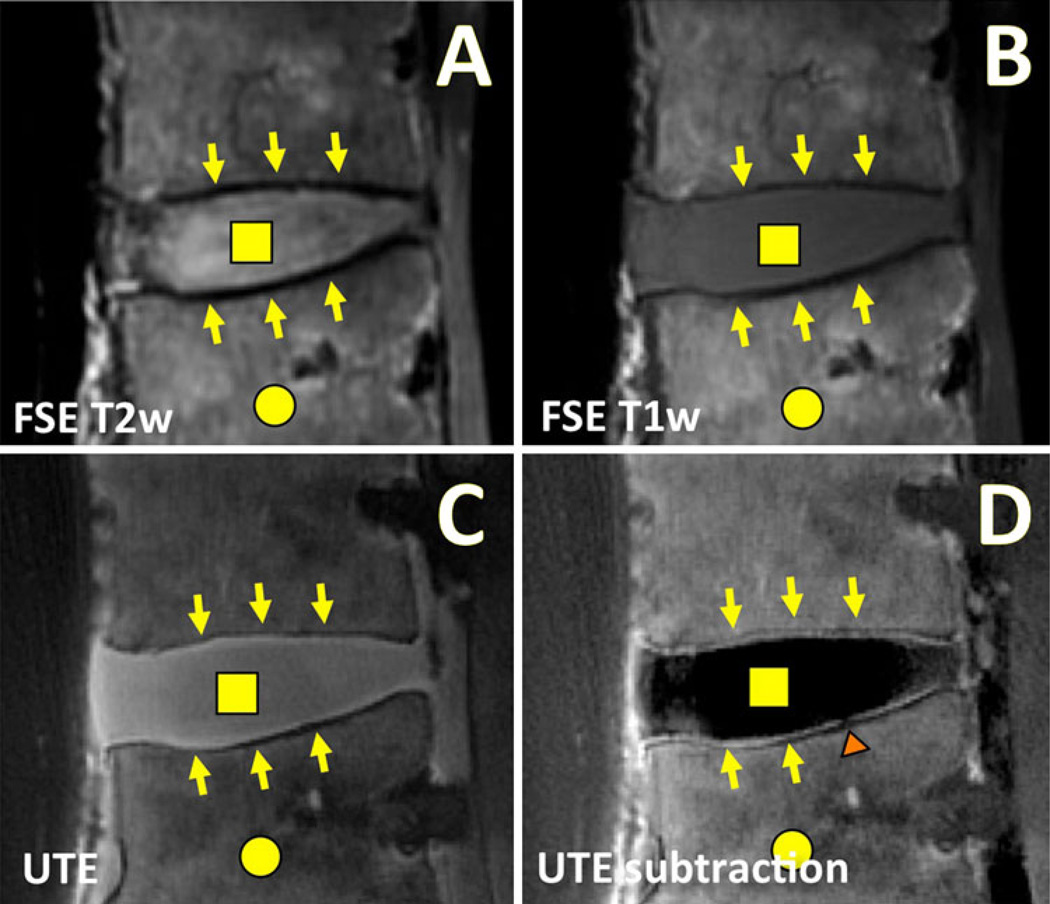
Morphologic MRI of a human lumbar spine in the sagittal plane using conventional and UTE techniques. Conventional fast spin echo (FSE) a T2-weighted (T2w; TR = 2,000 ms, TE = 80 ms) and b T1-weighted (T1w; TR = 600 ms, TE = 10 ms) MR images are often used for clinical evaluation of the disc proper (square) and the bone marrow (circle). These conventional MR images exhibit low signal intensity at the disco-vertebral junction (arrows). c UTE MR images obtained at TR = 300 ms and TE = 0.01 ms reveal high signal intensity at the disco-vertebral junction (arrows) as well as the disc proper (square). d After subtracting the second echo image (at TE = 11 ms) from the UTE image, the osteochondral junction is seen with a much greater contrast. A focal region of abnormal disco-vertebral junction (triangle) can now be seen

