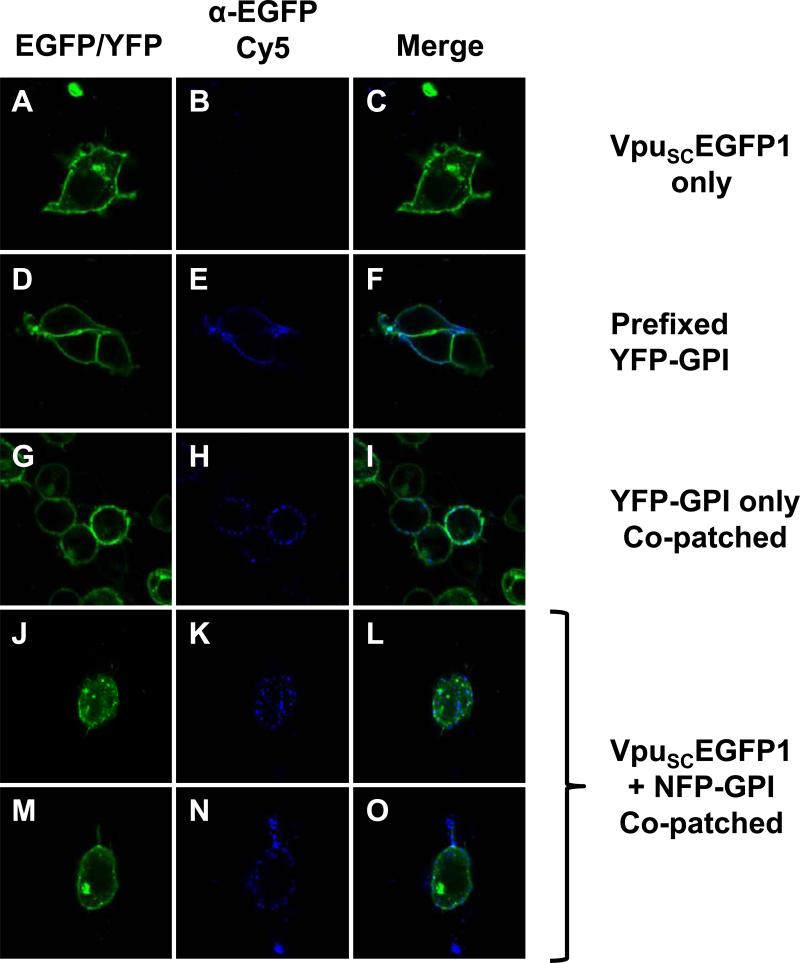
Figure 5.
Co-patching experiments reveal that VpuSCEGFP1 partially co-localizes with NFP-GPI. Panels A, D, G, J, and M are fluorescent micrographs using a filter for YFP/EGFP . Panels B, E, H, K, aand N are fluorescent micrographs for Cy5. Panels C, F, I, L, and O are a merge of the two fluorescent micrographs to the left. Panels A-C. 293 cells were transfected with vector expressing VpuSCEGFP1. Note the punctuate fluorescence in Panel C. Panels D-F. 293 cells were transfected with vector expressing YFP-GPI and pre-fixed. Panels G-I. 293 cells were transfected with a vector expressing YFP-GPI and co-patched. Panels J-O. 293 cells were transfected with both VpuSCEGFP1 and NFP-GPI.

