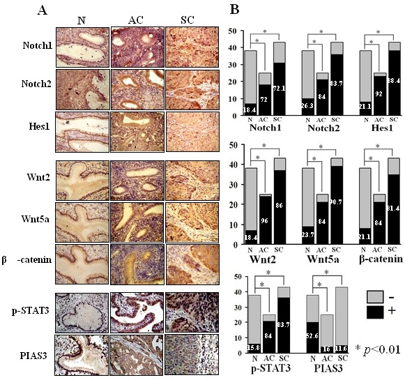
Figure 4. Frequencies of Notch1, Notch2, Wnt2 and Wnt5a detection and nuclear translocation of Hes1, β-catenin, p-STAT3 and PIAS3 in human cervical cancer specimens.

A: Tissue microarray-based immunohistochemical staining for Notch1, Notch2, Hes1, Wnt2, Wnt5a, β-catenin, p-STAT3 and PIAS3 in normal cervical tissues removed from the uterine fibroid patients at post-reproduction ages (N), cervical adenocarcinomas (AC) and cervical squamous cell carcinomas (SC) (original magnifications×400). B: Histogram of Table 1, *p<0.01.
