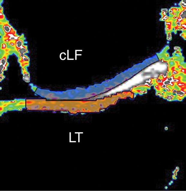
Figure 2a:
Representative ROIs for (a) lateral and (b) medial compartments of femorotibial joint used in analysis of cartilage T2. ROI was limited to center section of coronal multisection multiecho source images. Orange indicates tibial ROIs, and blue indicates femoral ROIs. cLF = central lateral femoral condyle, cMF = central (weight-bearing) medial femoral condyle, LT = lateral tibia, MT = medial tibia.

