
Figure 1.
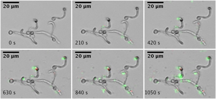
Real-time microscopic analysis of adhesion of A. actinomycetemcomitans to C. albicans SC5314. Hyphae of C. albicans SC5314 were allowed to form on the bottom of a microfluidics plate. Bacteria were stained with Syto9 and PI and allowed to adhere to C. albicans SC5314 while flowing at 0.5 dyne cm−2. Images were captured every 30 s for a total of 10 min (montage shows images every 210 s). Images were edited for brightness and contrast.