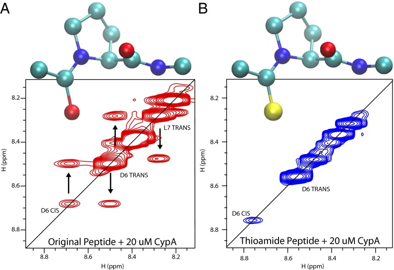
Fig. 5.
Homonuclear NOESY spectra of the model peptide (A) and thioamide-substituted peptide (B) in the presence of 20 μM cyclophilin A after a 200-ms mixing time. Exchange peaks, which indicate isomerization, are visible for the model peptide but not for the thioamide-substituted peptide. The sulfur atom is shown in yellow in B.

